
The Judi platform de-identifies and quality checks every trial image at the source, then delivers it the same day wherever your trial needs it: a core lab, an independent reader, an eligibility packet. Images arrive complete and fully analyzable, protecting the eligibility and endpoint decisions that depend on them.

Central imaging in clinical trials is how medical images, such as MRI, CT and PET scans, are acquired locally at investigator sites but analyzed centrally, by independent readers or an imaging core lab. One independent team evaluates every scan against the same criteria. A single workflow keeps the images consistent and analyzable, supporting eligibility determinations and endpoint outcomes. When a trial depends on image-based decisions, delays land on DSMB review schedules, interim results and database lock.
Judi supports central imaging in three areas: collection, processing and accessibility. Sites upload images online. Automated de-identification prevents PHI escape and masks references to treatment. Automated QC confirms the modality is correct, the imaging is complete and the images comply with the trial’s data acquisition guidelines, so no image-based decision is lost to an unanalyzable scan.
Judi then transmits images to a core lab, routes them to another destination such as a document repository, or holds them centrally for download by authorized parties. Every action is captured in a timestamped audit trail. The platform is SOC 2 Type 2 certified and aligned with 21 CFR Part 11, HIPAA and GDPR.
Clinical trial images pass through many hands: sites, CROs, AROs, independent reviewers, core labs and sponsors. Manual processes, couriers, email and separate trackers slow the images down and scatter the records, adding risk at every step. Trials get delayed, PHI gets exposed, endpoints are lost and the record gets harder to defend.
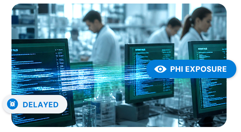
Obtain fully analyzable images on time, exactly where you want them.
Judi replaces manual processing silos with automated, trial-specific workflows to protect study timelines and data integrity.
Images arrive 100% analyzable. Automated upload checks immediately catch quality and modality errors at the source.
Uploads, de-identifications, QC actions and queries are captured as they happen in a timestamped audit trail.

Submit images through a browser with nothing to install.

Intake, processing and delivery configured to the trial’s imaging charter.

Trial-specific rules automatically remove targeted PHI from metadata, private and hidden DICOM tags.
Pixel-level PHI can be masked at the source, before the image enters the workflow.

Every upload, de-id and action timestamped and captured in real time to 21 CFR Part 11 standards.

Automatically transmit images to any destination the trial requires, or hold for authorized access.
Submit processed images with eligibility packets or endpoint dossiers.

Audit logs, transfer data and transmittal forms.

Almost everything in a trial gets a second chance. A form can be updated. Data can be corrected. A scan cannot. The image taken at week 12 exists only at week 12. When a reader opens it three weeks later and finds it unusable, the patient has gone home, the window has closed. Judi quality checks each image at upload so issues can be resolved before the patient leaves. Image quality and cycle times improve, your images are analyzable on receipt and your trial maintains momentum.
An over-scrubbed image is private and compliant, but worthless. Judi applies trial-specific mapping to remove or reassign only targeted identifiers. This preserves essential data, like acquisition sequences and the metadata central readers require. By applying fit-for-purpose de-identification at the source, Judi protects patient confidentiality and the study blind before images leave the local machine.
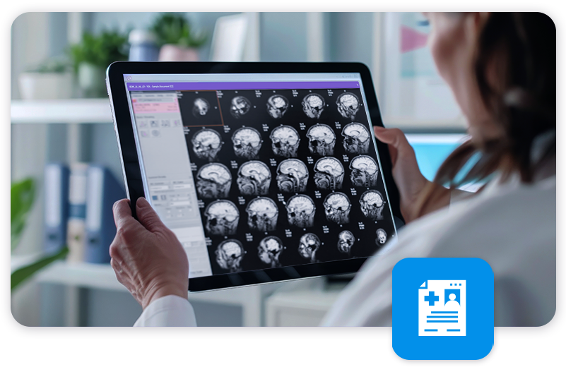
An imaging core lab is an independent body that centrally analyzes imaging data and provides standardized decisions, such as eligibility determinations or endpoint assessments. Unlike a central lab, which generates test results, a core lab uses images as the basis for its analysis, much as endpoint adjudication committees use source documents as the basis for theirs.
Your independent readers or your core lab perform the centralized imaging review after receiving complete, analyzable images from Judi. While it varies by protocol complexity, turnaround across our core lab partners typically ranges from 24 to 48 hours. Judi removes courier transit and catches quality errors at upload, taking the impact of manual processes out of the timeline.
De-identification runs on the local machine, before an image is uploaded. Judi uses a trial-specific schema to automatically remove or reassign the PHI in the DICOM tags, and the user can also mask anything burned into the pixels. Nothing leaves the source machine until de-identification is complete.
Patient identifiers and clinical parameters all occupy fields in the DICOM metadata, so indiscriminate scrubbing can strip the acquisition detail readers depend on. To prevent this, Judi uses trial-specific mapping to remove or reassign only PHI identifiers and any references to treatment. This keeps acquisition sequences and required metadata intact. Images arrive anonymous and fully analyzable.
Imaging protocol deviations occur when image acquisition does not comply with the trial’s image acquisition guidelines. These deviations are primarily technical: artifacts, reduced image quality, the wrong slice thickness, the wrong magnetic field strength, flipped angles, missing sequences or incorrect patient positioning. Many go unnoticed until the reader opens the file. By then it is too late to take preventive or corrective action, because the timepoint has passed, the images are unanalyzable and the endpoint is lost.
Unanalyzable scans are prevented by checking each image against the specified modality and the trial’s image acquisition guidelines at the moment it is uploaded, rather than somewhere down the line when a reader opens it. Judi runs automated quality checks at submission, so acquisition problems surface while the site can still act on them and the patient may still be on site. Catching a problem three weeks later, when the imaging window has closed, means a lost assessment and a lost endpoint.
Your independent reviewers or your core lab perform the blinded independent central review. Judi works seamlessly with core labs to handle the image collection, processing and accessibility workflow, and the readers handle the analysis. Judi supports blinded review by removing references to treatment or other identifiers that would unblind the reviewers, so detail that might bias a read does not travel with the images. Readers see only what they should.
Yes. Imaging data is collected, de-identified and transmitted in a secure, validated, access-controlled platform. Yes. Judi is SOC 2 Type 2 certified and holds active certification under the EU-U.S. Data Privacy Framework, including the UK Extension and the Swiss-U.S. DPF. The SOC 2 examination is an independent CPA review of the design and operating effectiveness of our security controls, performed every year since 2021, with no exceptions noted in the most recent report. Judi operates as a data processor and business associate on behalf of sponsors and CROs, and is designed and validated to support your 21 CFR Part 11, HIPAA and GDPR obligations, covering electronic records and signatures, audit trails and access controls. Imaging data is collected, de-identified and transmitted in a validated, access-controlled platform, with every action captured in a timestamped audit trail. AG Mednet supports client security questionnaires, vendor assessments and audits as part of standard engagement, and can share evidence on request.